Image segmentation is the process of dividing an image into regions. This process is problem-oriented. Examples of segmentation are illustrated using two types of images of the heart, cineangiocardiographic and a nuclear medicine images. In the first case, the challenge is to separate the blood (light) area from the rest. In the second case, the problem is to separare the live tissue (light) area from the rest.
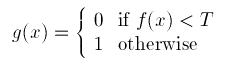
The simplest and most widely used segmentation method is thresholding. It consists of setting background values for pixels below a threshold value T and a different set values for the foreground.
In the experiment below, the variation on the threshold value causes a large variation on the area of the foreground pixels. This is a difficult problem to solve.
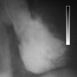
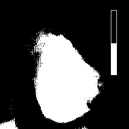
Shown below are the original image and the results after applying different thresholds values (118, 128, 138) to it.
 --
--

Original image ---------------- Threshold: 118
 --
--
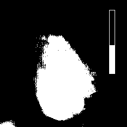
Threshold: 128 ---------------- Threshold: 138
The areas of each thresholded image are depicted below.
Level    Area
118     19,670 pixels
128     16,969 pixels
138     14,462 pixels
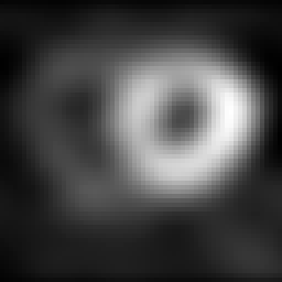
Depicted below are the original image and the results after applying different thresholds values (118, 128, 138) to it. The original image has been first expanded or "zoomed" by a factor of 4.
 --
--

Original image ---------------- Threshold: 118
 --
--

Threshold: 128 ---------------- Threshold: 138
The areas of each thresholded image are depicted below.
Level    Area
118      731 pixels
128      659 pixels
138      588 pixels